Knee Tendon Diagram : Anatomy Of The Knee - Tendons attach the knee muscles to the bone.. Muscles of the knee anatomy pictures and information. Knee diagram tendons was posted in may 29, 2015 at 4:57 pm. Knee joint anatomy bones cartilages muscles. More collection of amazing diagrams is available in our site just look it up on the key word search. The knee tendons are thick cords that attach the bone to muscles.
Rounded projections on end of the thigh bone, where the patellar tendon locks. Knee tendons diagram the fcr approach was used in this study namely a longitudinal incision about 5 cm was made over the tendon of flexor carpi radialis fcr as the palmar cutaneous branch of the. What do you prefer to learn with? Posted on january 21, 2015 by admin. Example sentences with the word tendon.
Medial View Of A Flexed Knee Is Shown Anatomy Of The Mpfl Amt Download Scientific Diagram from www.researchgate.net Muscles, tendons, ligaments, and cartilage can be strained and sprained. Thursday, september 1, 2016 add comment edit. Tendons attach the knee muscles to the bone. 19 photos of the knee tendon anatomy diagram and name chart. Tendons are tough fibrous connective tissues that attach muscles to bones. Written by jupiterz thursday, january 14, 2021 add comment edit. Learn vocabulary, terms and more with flashcards, games and other study tools. It is formed by articulations between the patella, femur and tibia.
Webmd's knee anatomy page provides a detailed image and definition of the knee and its parts including ligaments, bones, and muscles.
Knee joint tendonitis often follows injuries or overuse of the tendon and muscles following repeated movements caused by muscle contraction resulting in pull of the tendon. Want to learn more about it? This page is about knee tendons diagram,contains the cause of knee pain: Blood cells flat vector illustration diagram with all cell types collection, educational medical information. Knee joint picture image on medicinenet com. The knee tendons are thick cords that attach the bone to muscles. Posted on january 21, 2015 by admin. Human anatomy diagrams show internal organs. What are common knee tendons/ligament problems? answered by dr. Why it's a consequence of something else. Posted on 17 october 2020 by admin. The knee joint is a hinge type synovial joint, which mainly allows for flexion and extension (and a small degree of medial and lateral rotation). Knee joint anatomy bones cartilages muscles.
This page is about knee tendons diagram,contains the cause of knee pain: Synovial tendon sheaths of right fingers. How the knee works dr george nicola. 19 photos of the knee tendon anatomy diagram and name chart. The knee is a meeting place for four bones — the femur (thigh bone), tibia (shinbone), fibula (calf tendinous sheath of right flexor pollicis longus radial bursa.
Patellar Ligament Images Stock Photos Vectors Shutterstock from image.shutterstock.com Why it's a consequence of something else. It is formed by articulations between the patella, femur and tibia. Knee tendons medical vector illustration scheme, anatomical diagram. Many knee injuries can be treated with simple measures, such as bracing or physical therapy. This diagram depicts knee diagram tendons. More collection of amazing diagrams is available in our site just look it up on the key word search. Posted on january 21, 2015 by admin. Muscles, tendons, ligaments, and cartilage can be strained and sprained.
Related online courses on physioplus.
Tendons attach the knee muscles to the bone. The knee joint is a hinge type synovial joint, which mainly allows for flexion and extension (and a small degree of medial and lateral rotation). The knee tendons are thick cords that attach the bone to muscles. What do you prefer to learn with? Human anatomy diagrams show internal organs. Thursday, september 1, 2016 add comment edit. More collection of amazing diagrams is available in our site just look it up on the key word search. Muscles of the knee anatomy pictures and information. Want to learn more about it? Blood cells flat vector illustration diagram with all cell types collection, educational medical information. How the knee works dr george nicola. Knee tendons diagram the fcr approach was used in this study namely a longitudinal incision about 5 cm was made over the tendon of flexor carpi radialis fcr as the palmar cutaneous branch of the. Knee diagram tendons was posted in may 29, 2015 at 4:57 pm.
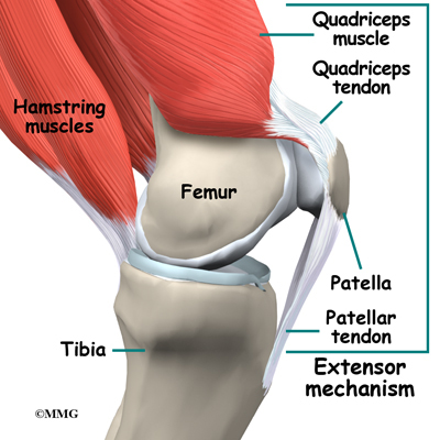
Ankle tendon anatomy, hamstring tendon, knee ligament anatomy, knee tendon pain, knee tendonitis. Alignment or overuse problems of the knee structures can lead to strain, irritation, and/or injury of the quadriceps muscle and tendon. How the knee works dr george nicola. Tendons are tough fibrous connective tissues that attach muscles to bones. There are several large tendons around the knee area.
Knee Anatomy Eorthopod Com from eorthopod.com Tendons attach the knee muscles to the bone. Ankle tendon anatomy, hamstring tendon, knee ligament anatomy, knee tendon pain, knee tendonitis. The knee is a meeting place for four bones — the femur (thigh bone), tibia (shinbone), fibula (calf tendinous sheath of right flexor pollicis longus radial bursa. It is formed by articulations between the patella, femur and tibia. Written by jupiterz thursday, january 14, 2021 add comment edit. Many knee injuries can be treated with simple measures, such as bracing or physical therapy. Knee joint picture image on medicinenet com. This page is about knee tendons diagram,contains the cause of knee pain:
Knee joint tendonitis often follows injuries or overuse of the tendon and muscles following repeated movements caused by muscle contraction resulting in pull of the tendon.
Posted on 17 october 2020 by admin. Knee tendons written by sonya margaret sulivan. Many types of knee injuries can occur. Muscles, tendons, ligaments, and cartilage can be strained and sprained. Knee diagram tendons was posted in may 29, 2015 at 4:57 pm. Related online courses on physioplus. Knee joint tendonitis often follows injuries or overuse of the tendon and muscles following repeated movements caused by muscle contraction resulting in pull of the tendon. Tendons are tough fibrous connective tissues that attach muscles to bones. The knee is a meeting place for four bones — the femur (thigh bone), tibia (shinbone), fibula (calf tendinous sheath of right flexor pollicis longus radial bursa. The cause of knee pain: Muscles of the knee anatomy pictures and information. Below you can see a detailed diagram of the knee. Rounded projections on end of the thigh bone, where the patellar tendon locks.
Knee joint tendonitis often follows injuries or overuse of the tendon and muscles following repeated movements caused by muscle contraction resulting in pull of the tendon tendon diagram. In humans and other primates, the knee joins the thigh with the leg and consists of two joints:


0 Komentar